Treatment of Cutaneous Atrophy After Corticosteroid Injection, With Heterologous Type I Collagen
Steroid atrophy often regresses over time, but patients are rarely patient enough to wait for spontaneous regression. Although the mechanism of atrophy after steroid injection is not yet fully understood, it is believed to result from a reduction in fibroblasts and collagen degeneration.
Steroid-induced atrophy is a well-documented complication of intralesional steroid use with an estimated incidence varying from 1.5% to 40%. Cutaneous atrophy usually begins within 2 to 3 months of injection and regresses within 1 to 2 years. However, it has also been reported to persist beyond 5 years.1 Treatment modalities that have been used to restore cutaneous atrophy include autologous fat injection, fat grafting, hyaluronic acid filler, pulsed dye laser, and intralesional bacteriostatic saline. We present 2 cases of steroid injection-induced atrophy treated with heterologous type I collagen (HTIC) (Linerase Collagen, Typeone Biomaterials Srl, Lecce, Italy).
Methods
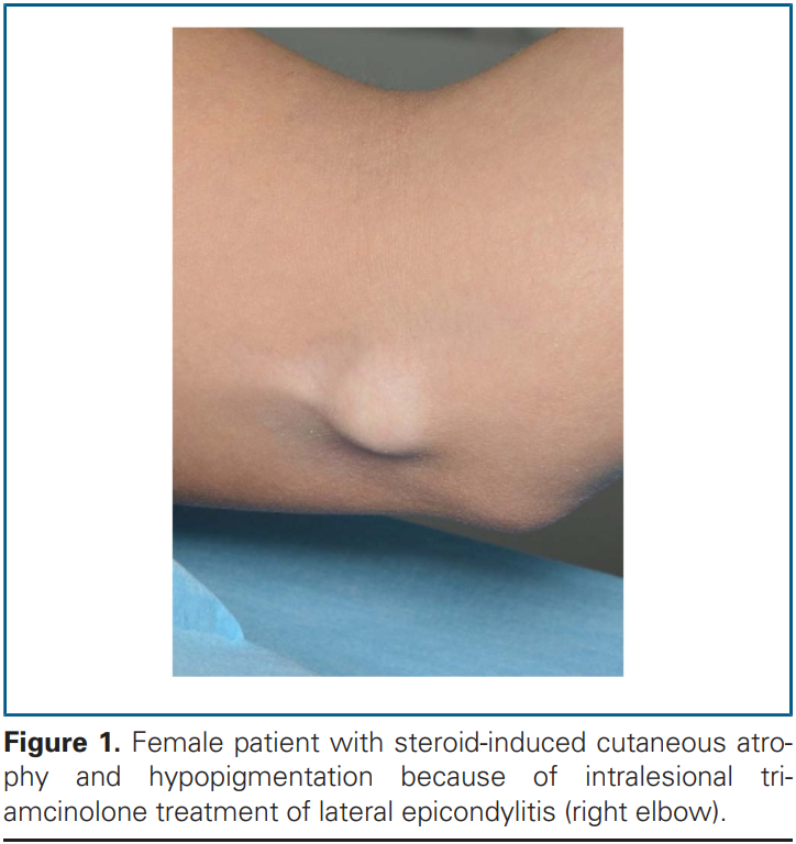
Two patients (1 male and 1 female) presented with corticosteroid-induced skin atrophy and hypopigmentation after steroid injection treatment. Clinical examination revealed 5- to 8-cm lesions that had not changed in size and had not been treated for at least 12 months. There were no signs of koebnerization and no history of keloids or hypertrophic scarring.
One patient was initially treated for lateral epicondylitis with local steroid injections (triamcinolone 10 mg), 3 injections in total, with an interval of 2 months between treatments. The patient experienced progressive skin atrophy, pigmentary changes, tenderness, and increased skin fragility after the third injection. The other patient had been treated for de Quervain’s tenosynovitis of the wrist with a methylprednisolone injection (40 mg) and showed cutaneous atrophy and hypopigmentation 2 to 3 months after the injection.
Xem thêm