Human Fibroblast attachment and growth into Ingenera Disposable System
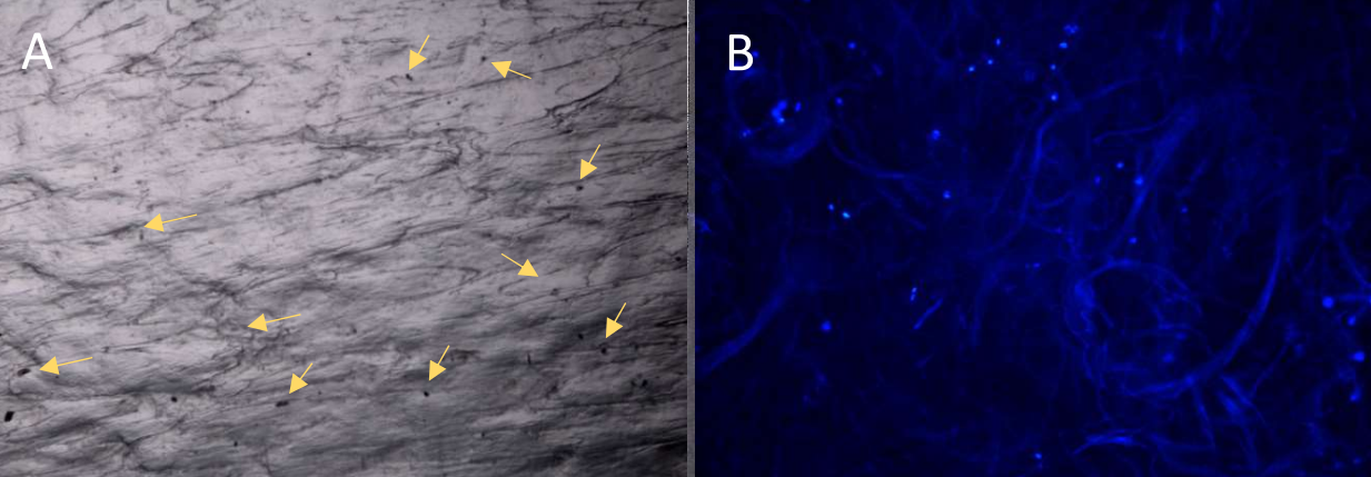
The aim of this study was to evaluate human fibroblasts attachment and growth into the disposable device INGENERA®. The results clearly demonstrated that human fibroblasts are able to adhere and grow into the device.
Type I collagen is the most abundant mammalian protein, present in the extracellular matrix of several tissues for which provides structural and mechanical scaffold.1,2 High biocompatibility and biodegradability make collagen ideal for biomedical applications3,4 since the fibrous collagen network facilitates cell adhesion/proliferation and cell migration to the wounded site, actively supporting wound healing.5 Collagen‐based dermal platforms have been applied to cover burn wounds, treat ulcers, reduce tissue contraction and scarring, and increase epithelialisation rate. 6‐10 The platforms have been shown to accelerate healing and wound closure favouring cell proliferation and tissue vascularization. Despite their rather extensive usage as biomaterials for scaffold design, collagen scaffolds remain sustainable materials with highly engineering potential.
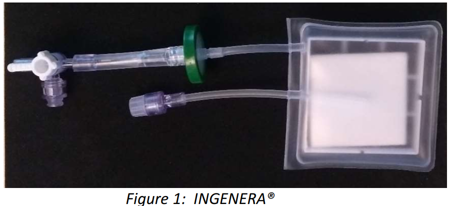
INGENERA® is a sterile disposable system made of a three‐dimensional plastic bag, equipped with tubes to allow the introduction into the bag of a suspension of skin cells and culture media, containing a flat sponge of native, not cross‐linked, lyophilized type I dermal collagen (Figure 1).
MATERIALS AND METHODS
Human primary fibroblasts from paediatric control were cultured in D‐Modified Eagle’s Medium (D‐MEM, Lonza) supplemented with 10% foetal bovine serum (FBS, Euroclone), 4 mM glutamine (Euroclone), 100 µg/ml penicillin and streptomycin (Euroclone) at 37°C in humidified atmosphere containing 5% CO2. After cells expansion, cells were trypsinized, counted and 5x10⁵ cells were plated in the collagen containing bags in the same growing media (25 ml). No growing media was added or substituted during the whole test. Collagen bags seeded with cells were incubated at 37°C in humidified atmosphere containing 5% CO2. Cells were used up to passage 7. Fibroblast’s attachment was evaluated 8 hours (h) after plating by cells counting with Biorad TC20 in presence of trypan blue (Biorad) to discriminate between live and dead cells.
Xem thêm